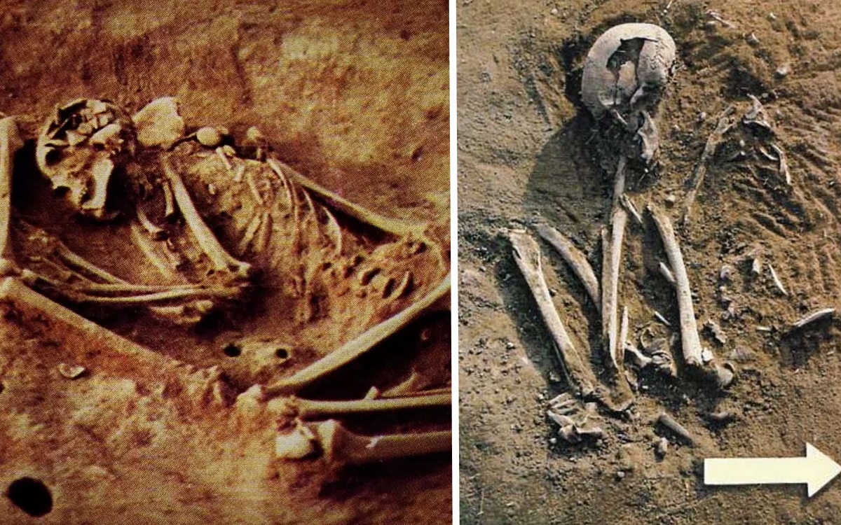
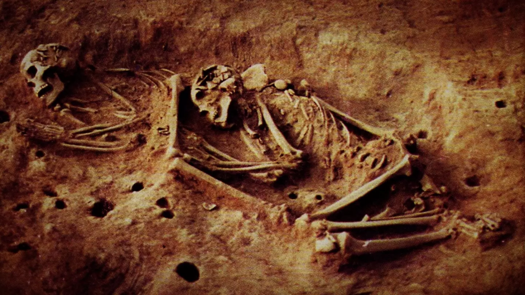
A team of archaeologists working in a remote valley of Eastern Anatolia has unearthed human remains dating back 6,000 years whose DNA patterns don’t match any known ancient population—raising the possibility that our understanding of early human migration and cultural exchange may need a radical rewrite. The discovery, announced this week in a study published in Nature, includes five skeletons found in a single burial pit, each exhibiting genetic markers previously unseen in Eurasian populations from that era.
Lead researcher Dr. Elif Demirci described the find in an interview with BBC News as “astonishing,” noting that while pottery styles and artifacts at the site align with the late Neolithic, the genetic signatures echo no known Anatolian or Mesopotamian lineage. “These individuals carry a genomic component unlike anything we’ve mapped in 10,000 years of ancient DNA research,” Demirci said.
“This discovery is a game-changer for our models of Neolithic settlement.” — Dr. Elif Demirci #AncientDNA— Archaeology News (@ArchNews) July 25, 2025
The burial site, located near the modern-day town of Van, was first surveyed in 2023 by the Istanbul University team collaborating with the Perseus Project. After clearing vegetation, they found pottery fragments decorated with geometric motifs similar to those of the Halaf culture, which thrived in northern Mesopotamia between 6,300 and 5,400 years ago. Yet when geneticists sequenced the ancient DNA, they were stunned to find mitochondrial haplogroups and autosomal components that align with populations now only seen in isolated North African desert communities, according to an analysis by the Harvard Reich Lab.
New paper reveals Anatolian Neolithics with North African lineage—did caravans cross the Sahara and Anatolia 6,000 years ago? #NeolithicMystery— Science News (@ScienceNews) July 25, 2025
If confirmed, the finding would suggest that prehistoric trade routes spanned far wider than previously thought, perhaps even connecting the Levant with Trans-Saharan corridors centuries before the rise of the Egyptian empire. Dr. Marcus Franks, a paleogeneticist at Max Planck Institute, applauded the study in Science Magazine, calling it “the strongest evidence yet that Neolithic societies were far more interconnected—and mobile—than our textbooks indicate.”
Archaeological context underscores the mystery. Alongside the skeletons, researchers found small bronze beads, obsidian arrowheads traced to sources in modern-day Armenia, and what appears to be a ceremonial staff carved from cedar—wood not native to Eastern Anatolia but commonly used in Phoenician shipbuilding, as detailed in a recent Journal of Near Eastern Studies article.
Phoenician-style cedar object found in Anatolia? The puzzle deepens. #PhoenicianArtefacts— Ancient World News (@AncientWorld) July 25, 2025
The unprecedented genetic profile was identified by comparing the new sequences against over 4,000 ancient genomes stored in the EVA database. While no exact match exists, the skeletons exhibit a 15–20% component resembling modern Tuareg and other Saharan groups, with the remainder similar to local Neolithic farmers. “It’s a genetic cocktail we’ve never documented in this region,” geneticist Dr. Sophia Rana explained to NPR’s Science Desk.
Public reaction underscores the discovery’s magnitude. On Twitter, #AncientConnections has trended internationally, with enthusiasts speculating about lost migrations, climate-driven exoduses, and forgotten civilizations. A popular thread by history blogger @LostVectors argued that a severe climatic event around 6,200 years ago—a rapid cooling known as the “6.2 ka event”—could have driven Saharan foragers toward river valleys in Anatolia, carrying their genes and crafts.
Could a sudden drought have driven Saharan nomads all the way to Anatolia? #AncientConnections— LostVectors (@LostVectors) July 26, 2025
Still, skeptics caution against overinterpretation. Dr. Elena Ortiz, an archaeologist at University College London, told The Guardian that “we need broader sampling across Eastern Mediterranean sites before rewriting migration maps. Anomalous DNA signals can arise from unrecognized local diversity as well.”
The team plans further excavations at the site, named Tell Kızıltepe, this fall, aiming to unearth additional graves and settlement layers. They also hope to perform isotopic analysis on the teeth of the skeletons to trace childhood diets and mobility patterns—a technique that proved illuminating in studies of Bronze Age nomads published by PNAS.
Meanwhile, the Peruvian government has offered to share their expertise in handling remote-field genetics labs, as they did for discoveries at Caral last year, according to a report by Peru This Week. The collaboration could streamline processing and help avoid contamination that plagued previous ancient DNA projects.
Whether these skeletons represent an unknown migrant wave or simply a local anomaly remains to be seen. But one thing is clear: as more ancient genomes are sequenced, the neat boundaries of prehistoric populations will continue to blur. The echoes of remote deserts may yet reveal pathways that connect distant peoples in ways our ancestors might never have imagined.